
The University of Wisconsin–Madison effort to launch a shared cryo-electron microscopy facility for the bioscience community is gathering momentum, with two new faculty hires and key technology investments this summer.
Tim Grant, a research specialist at the Howard Hughes Medical Institute’s (HHMI) Janelia Farm Research Campus, is the latest addition to the core team. Grant will be a virology investigator for the Morgridge Institute for Research and an assistant professor of biochemistry at UW–Madison when he comes to Madison in early 2020.
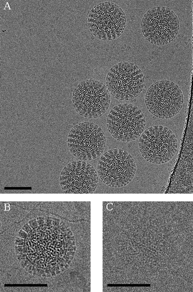
Cryo-electron microscopy (or cryo-EM, for short) is the latest essential tool for biologists trying to visualize and understand structures at the atomic scale. Cryo-EM flash-freezes biological samples at lightning speeds, without creating ice crystals that would warp the specimen. The result is a biological sample captured in its native state.
The process is helping scientists precisely define, at or near atomic resolution, the 3D structures of proteins, the large molecules that do most of the work in creating the body’s tissues and organs. By understanding proteins at this new, mechanistic level, scientists can begin to see how drugs interact with proteins, potentially leading to a revolution in creating new drugs and improving existing ones.
Grant’s first exposure to cryo-EM was in the early 2000s, as an undergraduate at Imperial College in London. Grant was drawn to the technology’s unique marriage of biology and computing, although the early imagery was primitive.
What would happen in the next 15-20 years is an explosion in resolution and image quality, to a point where the atomic nooks and crannies of proteins are becoming visible. Grant contributes to its broader use as developer of a software package called cisTEM, which places the complete workflow for cryo-EM analysis in a user-friendly interface for biologists.
Grant is motivated by the almost limitless potential of cryo-EM in impacting health. “Many diseases have a structural component that cryo-EM can peer into,” he says.
Grant’s current research challenge is better capturing the natural motion of these molecules. “A lot of important molecules, they really act as machines,” he says. “They move around. But the way standard processing works, when you have motion you also lose a lot of information. Finding a way to capture motion not only solves a problem, it adds a whole layer of extra information.”
Paul Ahlquist, director of the John W. and Jeanne M. Rowe Center for Research in Virology at Morgridge, says Grant will be an ideal talent for the initiative.
“We found a person who not only can both develop novel approaches that haven’t been pursued before, but also has the interest and ability to work with other collaborators to make sure those approaches get applied to important research problems.”
Paul Ahlquist, director of the John W. and Jeanne M. Rowe Center for Research in Virology at Morgridge
“We found a person who not only can both develop novel approaches that haven’t been pursued before, but also has the interest and ability to work with other collaborators to make sure those approaches get applied to important research problems,” Ahlquist says.
Along with Grant, the Institute for Molecular Virology and Department of Biochemistry are welcoming Assistant Professor Robert Kirchdoerfer in August. Kirchdoerfer uses cryo-EM to study the structure of virus components in order to better understand their biology and find targets for vaccines or antivirals. The Biochemistry Department is currently searching for one more faculty member who works in cryo-EM (Application information is here).
Several years ago, Ahlquist and others teamed with faculty leaders in biochemistry, including Biochemistry Chair Brian Fox, to make a campus-wide push for a major investment in cryo-EM. Groups contributing funding to the $15 million-plus initiative included Biochemistry, Morgridge, the School of Medicine and Public Health and its departments of biomolecular chemistry and neuroscience; and the Office of the Vice Chancellor for Graduate Research and Education.
In 2018, the group recruited Professor Elizabeth Wright to direct the UW–Madison Cryo-Electron Microscopy Research Center, which will house the majority of cryo-EM instrumentation on campus and be located in the Department of Biochemistry. It will serve as a research resource for all of campus as a research core. The new facility is expected to open in the spring or summer of 2020.
Construction is well underway in the Hector F. DeLuca Biochemical Sciences Complex for the facility, which will house four microscopes and other equipment in two buildings. The facility will house the powerful Titan Krios 300 kV transmission electron microscope (TEM). In the same building, a smaller 120 kV TEM will be located in the Biochemistry Optical Core (BOC) and Biophysical Instrumentation Facility (BIF) nearby.
“We thought about the value to the research community of bridging between the light microscopy and the biophysics resources of the BIF and BOC with the electron microscopy the cryo-EM facility,” Wright says. “It is important to bring researchers together to foster new ideas and collaborations, our having a microscope in BIF and BOC resource can be that nucleating factor.”
In the basement of the historic Hector F. DeLuca Biochemistry Building, the facility will have two more microscopes — a Talos Arctica 200 kV TEM and an Aquilos cryo-FIB-SEM — specimen prep equipment, and lab space for UW–Madison investigators, external collaborators and industry partners. The construction on phase two began in July 2019.
Construction and installation is complex because the rooms need temperature and humidity control and sophisticated shielding to make sure the microscopes are vibrationally isolated. Vibrations from nearby passing trains or elevators, as well as magnetic fields from other research equipment, can all interfere with the microscopes.
Ahlquist was also part of a second effort to bring an additional electron microscope to campus. Ahlquist, an HHMI investigator, teamed with two other HHMI investigators—Ed Chapman and Phil Newmark—to apply for high-end microscopy through a special HHMI fund. They received a $1.3 million grant, and were able to secure another $700,000 from campus partners to complete the purchase. The instrumentation will be complementary to others available to campus.
Even without the facilities open, the group has been spreading the word about cryo-EM, laying the groundwork for collaborations, and gathering preliminary data at other facilities. For example, they have started work with bacteriology professor Katrina Forest on altered bacterial membrane architecture, with bacteriology and biochemistry professor Bob Landick on transcription complexes, and with researchers at the School of Medicine and Public Health to develop vaccine candidates for different viruses. Another project involves scientists at the Great Lakes Bioenergy Research Center.
Wright has been working with Ahlquist and others to recruit a core team of faculty who heavily utilize cryo-EM in their work. Wright is also currently hiring a computer scientist/systems administrator staff member for the facility.
“It has been exciting to put together this team and we are thrilled to see it continue to grow,” Wright says. “When establishing a great cryo-EM community, it is important to recruit individuals with strengths in different biological areas, such as bacteriology, cell biology, and virology, combined with an interest in computation and technology development, so we can push the boundaries of the field at UW–Madison.”