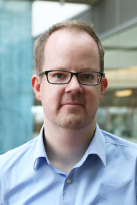
Affiliate, Morgridge Biomedical Imaging
Area:
Phone:
(608) 316-4417
Email:
[javascript protected email address]
Huisken has been awarded a prestigious Alexander von Humboldt Professorship from Germany, and in October 2021 accepted a research position at the University of Göttingen. Huisken remains an affiliate of the Morgridge Institute.
The overall goal of my lab is the systematic study of developmental processes in living organisms by noninvasive biomedical imaging techniques such as optical microscopy. Of primary interest is the investigation of organogenesis in zebrafish with special emphasis on the function and morphogenesis of the cardiovascular system and the endoderm. We develop novel quantitative microscopy tools and experimental strategies to understand and describe tissue dynamics on a cellular level. High-speed fluorescence microscopy is the primary tool to capture the dynamics of a heartbeat and the fate of single cells during organogenesis.
In our interdisciplinary lab we address all experimental steps from innovative transgenic lines and microscope development to systematic image processing. The biologists in the lab have the opportunity to use cutting edge microscopy to perform experiments that are impossible with off-the-shelf instruments. At the same time the physicists in the lab can build novel microscopes that are immediately applied to address exciting biological questions.
Areas of Expertise
- Selective plane illumination microscopy (SPIM)
- High-speed fluorescence microscopy
- Organogenesis
- Zebrafish, with special focus on the cardiovascular system and endoderm
Since 2016
Director of Morgridge Medical Engineering at Morgridge Institute for Research, Madison
Visiting Professor, Department of Integrative Biology
2010-2017
Research Group Leader at the Max Planck Institute of Molecular Cell Biology and Genetics
2005-2009
Postdoctoral work at the University of California, San Francisco
2004-2005
Postdoctoral work at the EMBL-Heidelberg
2000-2004
PhD work at the EMBL-Heidelberg, PhD in Physics, University of Freiburg
Selected Publications
- Image quality guided smart rotation improves coverage in microscopy
J. He, J. Huisken
Nature Communications 11, 150 (2020) - Multi-scale imaging and analysis identify pan-embryo cell dynamics of germlayer formation in zebrafish
G. Shah, K. Thierbach, B. Schmid, J. Waschke, A. Reade, M. Hlawitschka, I. Roeder, N. Scherf, J. Huisken
Nature Communications 10, 5753 (2019) - Putting Advanced Microscopy in the Hands of Biologists
R.M. Power, J. Huisken
Nature Methods 16, 1069 (2019) - Multi-sample SPIM image acquisition, processing and analysis of vascular growth in zebrafish
S. Daetwyler, U. Günther, C.D. Modes, K. Harrington, J. Huisken
Development 146, dev173757 (2019) - Dynamic, non-contact 3D sample rotation for microscopy
F. Berndt, G. Shah, J. Brugues, J. Huisken
Nature Communications 9, 5025 (2018) - Cell-accurate optical mapping across the entire developing heart
Michael Weber, Nico Scherf, Alexander M Meyer, Daniela Panáková, Peter Kohl, Jan Huisken
eLife 6 (2017), doi:10.7554/eLife.28307 - A guide to light-sheet fluorescence microscopy for multiscale imaging
Rory M Power, Jan Huisken
Nat Methods, 14(4) 360-373 (2017), doi:10.1038/nmeth.4224 - Hyperspectral light sheet microscopy
Wiebke Jahr, Benjamin Schmid, Christopher Schmied, Florian Fahrbach, Jan Huisken
Nat Commun, 6 Art. No. 7990 (2015), doi:10.1038/ncomms899 - The smart and gentle microscope
Nico Scherf, Jan Huisken
Nat Biotechnol, 33(8) 815-818 (2015), doi:10.1038/nbt.3310 - Optical tomography complements light sheet microscopy for in toto imaging of zebrafish development
Andrea Bassi, Benjamin Schmid, Jan Huisken
Development, 142(5) 1016-1020 (2015), doi: 10.1242/dev.116970 - High-resolution reconstruction of the beating zebrafish heart
Michaela Mickoleit, Benjamin Schmid, Michael Weber, Florian Fahrbach, Sonja Hombach, Sven Reischauer, Jan Huisken
Nat Methods, 11(9) 919-922 (2014) , doi:10.1038/nmeth.3037 - High-speed panoramic light-sheet microscopy reveals global endodermal cell dynamics
Benjamin Schmid, Gopi Shah, Nico Scherf, Michael Weber, Konstantin Thierbach, Claudia Campos, Ingo Roeder, Pia Aanstad, Jan Huisken
Nat Commun, 4 Art. No. 2207 (2013), doi:10.1038/ncomms3207 - Multilayer mounting enables long-term imaging of zebrafish development in a light sheet microscope
Anna Kaufmann, Michaela Mickoleit, Michael Weber, Jan Huisken
Development, 139(17) 3242-3247 (2012), doi: 10.1242/dev.082586 - Optogenetic control of cardiac function
Aristides B. Arrenberg, Didier Y.R. Stainier, Herwig Baier, Jan Huisken
Science, 330(6006) 971-974 (2010), DOI: 10.1126/science.1195929 - Arterial-venous segregation by selective cell sprouting: an alternative mode of blood vessel formation
Shane P Herbert, Jan Huisken, Tyson N Kim, Morri E Feldman, Benjamin T Houseman, Rong A Wang, Kevan M Shokat, Didier Y.R. Stainier
Science, 326(5950) 294-298 (2009), doi: 10.1126/science.1178577 - Selective plane illumination microscopy techniques in developmental biology
Jan Huisken, Didier Y.R. Stainier
Development, 136(12) 1963-1975 (2009), doi: 10.1242/dev.022426 - Even fluorescence excitation by multidirectional selective plane illumination microscopy (mSPIM)
Jan Huisken, Didier Y.R. Stainier
Opt Lett, 32(17) 2608-2610 (2007), https://doi.org/10.1364/OL.32.002608 - Optical sectioning deep inside live embryos by selective plane illumination microscopy
Jan Huisken, Jim Swoger, Filippo Del Bene, Jochen Wittbrodt, Ernst H K Stelzer
Science, 305(5686) 1007-1009 (2004), DOI: 10.1126/science.1100035
